Quantitative Photoacoustic Elastography
Photoacoustic imaging is a new biomedical imaging modality based on the use of laser-generated ultrasound, where tissue is illuminated by a short (∼ ns) laser pulse. The absorption of its light leads to a pressure wave via the photoacoustic effect, which is then recorded by an array of sensors. These data can be used to reconstruct the initial pressure distribution. It combines the high-contrast and spectroscopic-based specificity of optical imaging with the high spatial resolution of ultrasound imaging. The resulting volumetric images are defined by sharp boundaries and homogeneous regions.
Elastography is implemented by applying a mechanical force to a specimen and visualizing the resulting displacement. It is used for visualizing elastic properties of a specimen or probe. Clinical applications include the detection of skin, breast and prostate cancer, detection of liver cirrhosis, and characterization of artherosclerotic plaque in vascular imaging [1]. Elastography is an on top imaging method: Basically, every imaging technique can be used as basis for elastographic imaging, such as ultrasound imaging, magnetic resonance imaging or optical coherence tomography. With all these techniques, it is possible to visualize momentum images, from which mechanical displacements μ can be calculated, which forms the basis of clinical examinations. For motion estimation in common techniques are optical flow and motion tracking algorithms. In ultrasound Elastography and optical coherence Elastography, these are specifically referred to as speckle tracking algorithm. Photoacoustics has as yet not been used as underlying imaging technique for elastography, but only as supplement to enhance contrast in US-elastography. This can be attributed to the weak speckle patterns in photoacoustics, which generally is considered advantageous for imaging, but makes it less suited for speckle tracking algorithms.
Recently it has been shown by our group [2] that an artificial texture pattern can be introduced by simulating the effect of a band-limited part of the measurement data on the imaging data. In this way the artificial speckle patterns improve the deformation estimation with generic speckle tracking algorithms.
The applicability of this technique to real photoacoustic tomography is made possible by the joint project with the Center of Medical Physics and Biomedical Engineering, where a Fabry Perot (FP) polymer film ultrasound sensor tomography system is available.
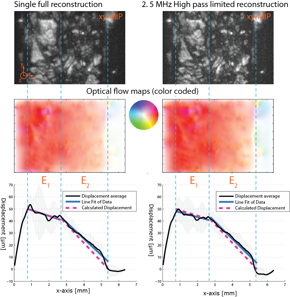
In a first step a proof of principle experiment is conducted [3]. Phantoms, which consist of Agar with ink inclusions, are compressed laterally, from the left side, along the x-axis by 50 &um;m. Pre- and post compression reconstructions are then used to determine the displacement via optical flow analysis. The Agar slabs have a predefined homogeneous elasticity indicated by the dashed blue lines in the figure. Thus the expected displacement curve can be calculated and compared to the measurement.
This ongoing work is funded by the Austrian Science Fund (FWF) within the Project P26687 - “Interdisciplinary Coupled Physic Imaging”.
References:
- [1] M. M. Doyley; Model-based elastography: a survey of approaches to the inverse elasticity problem. Phys. Med. Biol., 57:R35–R73, 2012.
- [2] T. Glatz, O. Scherzer, and T. Widlak. Texture generation for photoacoustic elastography. J. Math. Imaging Vision, 2015. Funded by the Austrian Science Fund (FWF) within the FSP S105 - “Photoacoustic Imaging”. Funded by the Austrian Science Fund (FWF) within the FSP P26687 - “Interdisciplinary Coupled Physic Imaging”.
- [3] J. Schmid, B. Zabihian, T. Widlak, T. Glatz, M. Liu, W. Drexler, W. and O. Scherzer. Texture generation in compressional photoacoustic elastography. Proceedings of SPIE, Photons Plus Ultrasound: Imaging and Sensing, Vol. 9323, 2015.